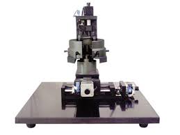
A great project for kids of any age is making a variety of microscope slides to view under your high power microscope.
When making temporary slides, the supplies needed are very basic:
1. blank slide
2. cover slip
In order to make your slide, simply place the specimen on the slide, apply the cover slip so it lays flat, and view under the microscope.
3. When finished viewing, wash the slide in warm soapy water. The cover slip is quite thin so be careful that you don't break it during cleaning.
Creating permanent slides involves sealing your specimen within the slide so it lasts, and then labeling the slide so that years from now you remember what the sample was.
You may also want to stain the slide, if so, this page offers great information on staining microscope slides.
1. Prepare the slide just as you would when making temporary slides.
2. When applying the cover slip, you will want to coat the very outer edges of the cover slip with a thin layer of clear nail polish or cement in order to permanently seal the cover slip to the slide.
3. Make sure your layer of adhesive is not too thick, as this will cause problems with not being able to focus properly on your specimen.
Fun slide making ideas!
1. Cheek cells from inside your mouth (use a Q-tip to scrape the inside of your mouth).
2. Pond water, or water from a muddy puddle.
3. Fine sand particles, sugar, salt, baking soda, a vitamin C tablet - any fine powdery substance. (If you have a polarizing filter - use it when viewing these items as well!)
4. Fabric fibers or strings.
Cotton - a thin strand pulled off a cotton ball.
5. Soil from the back yard.
A blade of grass or a thin section of a flower.
Tuesday, May 28, 2013
Thursday, May 23, 2013
25 Amazing Images for an Electron Microscope
Check out the top 25 most amazing images taken with a high powered electron microscope.
(Via list25 Youtube Channel)
Wednesday, May 22, 2013
Micro Empire
Clemens Wirth & Radium Audio presents: Micro Empire.
Moving on from Macro Kingdom, we pass through the portal of a microscope to venture into the Micro Empire. Molecular conflict and mitochondrial warfare … a heartstopping, subcellular epic … a truly microcinematic experience.
Clemens says, “As an enthusiast for little things, I wanted to go deeper than the macro universe, so I found myself hanging on the eyepiece of a microscope. The real challenge was definitely the small depth of field in microscopy. It’s really fascinating how detailed this tiny world is.”
Monday, May 20, 2013
Types of Microscopy
Almost everyone at some point in time has operated a compound microscope in the classroom to examine specimens and slides. Some children who opt for the home school route even have compound microscopes of their own. It is hard to argue the fact that microscopes are fundamental observation tools for earth science, biology, and other academic field such as chemistry Microscopes allow us to observe small details about specimens that would not be visible to the naked eye. The most commonly used forms of microscopy are electron microscopy, optical microscopy, and scanning probe microscopy.
Electron Microscope
Electron Microscopy requires a different variation of light and can achieve up to 10,000,000 X magnification level. They have revolutionized microscopy and our viewing of the nature around us.

Optical Microscopy
The most widely used form of microscopy and the beginning tool for observing the natural world. Optical microscopy is still in wide use today and has allowed us to observe bacteria and identify illnesses.
Scanning Probe Microscopy
Developed in the late 1980's this method is based on quantum mechanics. It is the highest level of microscopy even making single atoms visible.

Electron Microscope
Electron Microscopy requires a different variation of light and can achieve up to 10,000,000 X magnification level. They have revolutionized microscopy and our viewing of the nature around us.

The most widely used form of microscopy and the beginning tool for observing the natural world. Optical microscopy is still in wide use today and has allowed us to observe bacteria and identify illnesses.
Scanning Probe Microscopy
Developed in the late 1980's this method is based on quantum mechanics. It is the highest level of microscopy even making single atoms visible.
Friday, May 17, 2013
Adjusting Condensers
The condenser aperture diaphragm is used to control the contrast and resolution of an image.
An improper setting of the condenser aperture diaphragm can be the cause of much frustration both for teachers and students.
Students may attempt to find the focus with the condenser aperture diaphragm all the way open.
Remember, an open condenser aperture diaphragm results in a low depth of field.
Students may not see anything at all when working with high magnifications because the image is too dark. In this case the diaphragm is closed too much. The diaphragm should not be used to control the amount of light, but for some specimens or magnifications there may simply be no way around this especially if the lamp is not very powerful. The higher the magnification, the more need for a condenser.
 Tips:
Tips:


- Completely close the condenser aperture diaphragm when starting to use the microscope.
- Rotate the low power objective into position and find the focus with the coarse focus knob. The larger depth of field and higher contrast makes it easier for the students to focus the specimen.
- When switching to a higher magnification, the students should start to gradually open the condenser aperture diaphragm, to observe the differences in image quality.
- Adjust the light intensity with the dimmer to prevent glare. Students should be made aware that the condenser aperture diaphragm should be adjusted to the numerical aperture value which is printed on the objective.
- Opening the diaphragm further will not increase image quality, but may result in glare. If the sample is thick, strongly stained or pigmented then the diaphragm has to be opened to allow more light to pass through the specimen. As a consequence, the depth of field becomes smaller.
- Use the fine focus adjustment knob to focus through the different layers of the specimen

Shop Darkfield Condensers here: http://bit.ly/16rya5k
Wednesday, May 15, 2013
Gem Microscopes
A gem microscope is similar to a biological or medical microscope in that it is binocular, and uses compound lenses.
A binocular magnifying device has two eyepieces so that both eyes are used at once. This is ideal for getting a good three dimensional view. In a compound scope, there is a set of lenses close to the object being magnified and a set in the eyepieces.
With this set-up, magnification is compounded, meaning that, for example, if the objective lens is 5x and the ocular lens is 10x, the total magnification is 50X (5X x 10X).
Gem scopes differ from biological scopes in that the total maximum magnification is usually lower and there are more lighting options.
Professional grade gem microscopes generally include:
- brightfield illumination
- darkfield illumination
- oblique fighting
- overhead lighting
- light diffusing system
- a system for immersing the object in liquid in a well for viewing
- polarizing lighting
- pivoting stone holder
Shop our selection of Jewel Gem Microscopes here.
Check out our Professional Gem Jewel Darkfield Stereo Microscope 12X-75X
On sale now for $1,244.99
Monday, May 13, 2013
Gemology: Diamonds
Diamonds are one of our links between the world back then and the world now. We don't know if they truly are forever but we do know that they have been in existence a very long time.
Diamonds are made of carbon atoms that are formed into a tightly packed structure with extreme heat, pressure, and oxygen conditions. Most diamonds form in the earth's crust or mantle but some deeper down. Under the earth's crust they form as impact diamonds under the deep earth. due to mass pressure. The redox reaction of CO2 or Methane forms diamonds. The complex pattern of diamonds could take anywhere from 3 billion years to form in the earth's mantle and brought so the earth's surface through magma.
Friday, May 10, 2013
Home School Microscope Projects: Crystals
This is a great project for homeschooling because it's a fun and simple observation project that will allow students to identify various crystal shapes under a microscope. Crystal patterns come in all sorts of shapes and sizes.
Supplies needed are:

Supplies needed are:
- Microscope
- Glass Slides
- Salts, Sugar, Lemon Juice
- Polarizing Film
Procedure:
1. Dissolve salt in water to create saltwater. Repeat this for other minerals you want to test. Put mixtures in clean glass slide.
2. Let slides dry overnight until the liquid is evaporated.
3. Observe formations at various magnification powers. Observe images in observation log.
4. If formations are difficult to observe, you can use polarizing film. Place one piece over the light source and place the second piece over the slide on top of the stage. Adjust intensity of light illumination as needed.
5. Observe the magnification at various powers, the film should make the crystals appear with different colors.
6. Record all changes and observations.

Wednesday, May 8, 2013
Darkfield Imagery Tips
Darkfield microscopy is a very simple method for viewing unstained specimens clearly because it is clearly visible. Good candidates for darkfield observation usually have a transparent background that way the light an illuminate the specimen and darken the surrounding area, making the image quality clearer and more intense. Good specimens to use include aquatic organisms, cells, and cultures.
To achieve good darkfield images make sure that the central light rays along the optical axis are blocked. Blocking these light rays allows for just oblique rays to pass through at sharp angles, striking the specimen and illuminating the image. A darkfield condenser will get you the results your looking for with your microscope. The condenser reflects a single surface that provide more even illumination.
We recommend the OMAX Compound Binocular Microscope with Darkfield Condenser and 5.0 MP Digital Camera for $549.99
Model No: M827-A191-C50
Monday, May 6, 2013
Compound Microscopes 101
A compound microscope can be used for medical research to a day at the beach. A compound microscope includes an eyepiece, stage clips, and objectives with different lenses, adjustable knobs, a power button, and a stage with light source. Other parts include the body, nose piece, arm, and stage stop.
It can be used in various science fields such as Microbiology and Geology. Forensic investigators and scientists identify crystal shapes to reference pharmaceuticals and other medical needs. Microscopes can also help them see human cells, minerals, and metals.
The setup of the optical parts plays an important ole in working with a compound microscope. The condenser focuses light onto the specimen with either bright-field or darkfield condenser attachments.
By understanding these basics, you can independently carry out an experiment for observing minute objects of your interest, and try to explore some of the incredible things with the help of this wonderful instrument. Nowadays, compound microscopes are used in various domains, within or outside science. Care should be taken while buying any of the microscopes, be it compound or digital microscope. For better understanding, you can always discuss with a science teacher or an expert.
Friday, May 3, 2013
Multi Viewing Attatchments
A multi-viewing attachment is one of the most beneficial accessories for your microscope if you are interested in group research and observations. Most multi-viewing microscopes are designed to allow for anywhere from two to six viewers. Over the years consumers wanted more heads for larger conferences and research and also for teaching purposes. Nowadays there are systems with even up to ten to a dozen heads, while they function properly, the image quality is not the greatest with so many heads. This is because any microscope with more then six heads will make the light not be evenly distributed The light gets harder to find and all the users may not be seeing the same quality image.
For teaching purposes, two or more persons to view the specimen simultaneously is a great feature. It is also useful got presentations. We recommend out dual binocular model which is great for labs, demonstrations, clinics, universities, and schools. Shop double binocular head compound microscope here.
Features Include:
-4 levels of magnification
-Dual binocular heads
-2 pairs of eyepieces
-4 achromatic objectives
-Variable intensity illumination

Wednesday, May 1, 2013
What is Phase Contrast Microscopy?
Phase contrast is a microscopy method used by Fritz Zernike. Zernike made multiple discoveries including the direct path of light. Phase contrast microscopy makes images appear darker against a light background. Phase contrast uses a condenser system and an objective lens. Phase is only useful on images that are colorless or transparent because it makes it difficult to see it's surroundings. Objects suitable for phase contrast microscopy are bacteria, protozoa, and cells. Zernike was awarded the Nobel Peace Prize in 1953 for his discoveries in Physics.
We recommend our 2000X Phase Contrast Compound LED Microscope with 9.0 MP Camera.
Shop here: http://bit.ly/14WLZrU
We recommend our 2000X Phase Contrast Compound LED Microscope with 9.0 MP Camera.
Shop here: http://bit.ly/14WLZrU
Product Features Include:
- High Quality Glass Elements
- High Definition Images
- Eight Levels of Magnification
- USB Camera
- Advanced Software
- Abbe Condenser
Subscribe to:
Comments (Atom)


